

lecture given By Dr. Alaa Khamees , Written By Prof. Khalil AL-SALEM
Amblyopia, commonly called a lazy eye, is one of the most frequent causes of reduced vision in children. Fortunately, many children improve when the condition is detected early and treated correctly. However, not every child with reduced vision has simple amblyopia. Sometimes, a child receives glasses and appropriate amblyopia treatment, yet the vision remains unexpectedly poor. This is sometimes described clinically as recalcitrant amblyopia or amblyopia that is not responding as expected. In these cases, the ophthalmologist should reconsider the diagnosis. The problem may be more than amblyopia. An underlying retinal, optic nerve, neurological, or developmental condition may be limiting the child’s vision. In our clinical practice, several children referred with difficult or resistant amblyopia were eventually found to have important underlying causes. These included optic nerve glioma, myelinated retinal nerve fibers, retinoschisis, ocular albinism, and significant high refractive error
When a child has what appears to be recalcitrant amblyopia, we should ask an important question: Is this truly amblyopia, or are we missing an underlying organic cause? I would like to share five cases from clinical practice where persistent poor vision led us to look again.
What Is Recalcitrant Amblyopia?
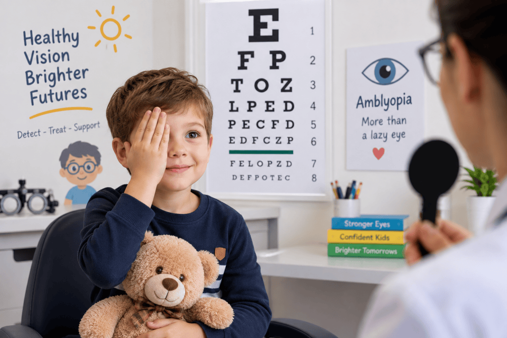
Amblyopia is reduced vision caused by abnormal visual development during childhood. It commonly results from strabismus, unequal refractive error between the two eyes, high refractive error, or obstruction of vision during early childhood. Importantly, true amblyopia is not primarily caused by structural disease of the retina or optic nerve. Treatment usually starts by correcting the refractive error. Some children also require patching of the stronger eye or other forms of amblyopia treatment. Improvement may take time. Therefore, slow improvement does not necessarily mean that the original diagnosis was incorrect. However, persistent poor vision despite appropriate treatment should raise an important question:
Is there another reason why this child cannot see better?
A careful reassessment may reveal a condition that was subtle or difficult to recognize during the initial examination.
Case 1: Optic Nerve Glioma Mistaken for Recalcitrant Amblyopia
One of our most important cases involved a child initially believed to have amblyopia. The poor vision did not respond as expected to treatment. Further evaluation eventually revealed an optic nerve glioma, also referred to within the broader group of optic pathway gliomas. Optic pathway gliomas occur predominantly in children. They can involve the optic nerve, optic chiasm, or other parts of the visual pathway. Reduced vision can be an early presentation. Other findings can include strabismus, proptosis, an abnormal pupillary response, optic disc changes, nystagmus, or visual field abnormalities. This case demonstrates why unexplained visual loss in a child should never automatically be attributed to amblyopia. If the visual loss is progressive, unusually severe, or inconsistent with the ocular examination, further investigation may be necessary. In selected children, this may include MRI of the brain and orbits.
Case 2: Myelinated Nerve Fibers and High Refractive Error
Another child had persistent poor vision associated with myelinated retinal nerve fibers. Myelinated retinal nerve fibers have a characteristic white, feather-like appearance during retinal examination. They may be associated with myopia, anisometropia, strabismus, and amblyopia. A particular combination of myelinated retinal nerve fibers, significant myopia, and amblyopia is often referred to as Straatsma syndrome. These cases can respond less successfully to conventional amblyopia treatment than uncomplicated refractive amblyopia. This does not mean that treatment should be abandoned. Accurate cycloplegic refraction, proper glasses, and appropriate amblyopia therapy remain important. However, parents should understand that an associated structural abnormality can limit the final visual outcome.
High Refractive Error Can Also Cause Amblyopia
High refractive error itself is an important cause of amblyopia. A child does not need to complain about blurred vision. Children often adapt remarkably well, particularly when they have had poor vision from an early age. This is why accurate refraction is essential.
A comprehensive assessment of suspected amblyopia should include visual acuity testing appropriate for the child’s age, ocular alignment assessment, cycloplegic refraction, pupillary examination, and examination of the anterior and posterior segments of the eye. Sometimes, repeating the refraction is worthwhile when the visual response does not match expectations.
Case 3: Retinoschisis Detected by OCT
Our third case demonstrates the importance of examining the macula carefully. The child had reduced vision that appeared to be amblyopic. However, the response to conventional treatment was disappointing. Further retinal assessment and optical coherence tomography (OCT) revealed schisis involving the foveal region. Retinoschisis causes splitting within the retinal layers. When the central retina or fovea is involved, central vision can be reduced. Children with retinoschisis may still have an associated amblyopic component. Therefore, appropriate refractive and amblyopia management may still be required. However, patching cannot correct the underlying retinal abnormality. This case illustrates the value of macular OCT in unexplained poor vision in children. Modern OCT can reveal subtle abnormalities that may not be obvious during routine fundus examination, especially in young children.
Case 4: Ocular Albinism and Foveal Hypoplasia
Another child initially treated for poor vision was eventually recognized as having features of albinism. Ocular manifestations of albinism can include reduced pigmentation, iris transillumination, nystagmus, refractive error, strabismus, and abnormal development of the fovea.
One of the most important causes of reduced vision is foveal hypoplasia. OCT can demonstrate the abnormal foveal development, including absence or reduction of the normal foveal pit. Some children have obvious features of albinism. In others, particularly those with less marked pigmentation changes, the diagnosis may be more subtle. These children may also have genuine amblyopia due to refractive error or strabismus. Treating that component is worthwhile. However, treatment of amblyopia cannot eliminate the visual limitation produced by significant foveal hypoplasia. Recognizing the diagnosis therefore helps us establish realistic expectations for the child and family.
When Should We Suspect That It Is More Than Amblyopia?
A child whose vision does not improve immediately should not automatically undergo extensive investigations. Amblyopia treatment requires time, and response differs between children. Nevertheless, certain findings deserve closer attention. Progressive visual loss is particularly important. So is vision that is much poorer than expected from the degree of strabismus or refractive error. An abnormal pupil, optic disc pallor or swelling, unusual nystagmus, visual field abnormalities, neurological symptoms, or an abnormal retinal appearance should also prompt further evaluation. Even when the fundus appears relatively normal, unexplained poor central vision may justify macular OCT or other investigations. Current ophthalmic guidance similarly emphasizes further investigation when the cause of visual loss remains unclear or visual function continues to deteriorate despite treatment. Depending on the clinical situation, this may include OCT, retinal imaging, electrophysiology, or neuroimaging.
What Should Be Rechecked When Amblyopia Treatment Fails?
When a child appears to have recalcitrant amblyopia, the first step is not necessarily to increase the hours of patching. The diagnosis should be reviewed. Visual acuity should be confirmed using an age-appropriate method. Refraction should be checked, preferably under adequate cycloplegia. Compliance with glasses and prescribed treatment should also be assessed. The ophthalmologist should then return to the clinical examination.
Are the pupils symmetrical?
Does the optic nerve look normal?
Is there subtle optic disc pallor?
Is the macula normal?
Could there be foveal hypoplasia or retinoschisis?
Is there unexplained nystagmus?
Is the visual loss compatible with the amount of refractive error or strabismus?
These questions can be more valuable than simply prescribing additional patching.
OCT Has Changed the Evaluation of Unexplained Poor Vision
OCT has become particularly useful in children with unexplained visual loss. It provides detailed cross-sectional images of the retina and macula without touching the eye. In our cases, OCT was especially valuable in identifying subtle macular pathology such as foveoschisis or retinoschisis and in demonstrating abnormal foveal development associated with ocular albinism. OCT does not replace careful examination. Rather, it adds another layer of information when the clinical findings and visual acuity do not fit together.
The Most Important Lesson From Recalcitrant Amblyopia
Amblyopia is common, and most children with amblyopia do not have serious underlying disease. Nevertheless, the diagnosis of amblyopia should not prevent us from reconsidering the child when treatment fails. Our experience with children eventually diagnosed with optic nerve glioma, myelinated retinal nerve fibers, retinoschisis, ocular albinism, and high refractive error demonstrates an important clinical principle. When the visual response does not fit the diagnosis, examine the child again. Sometimes the child truly has difficult amblyopia. Sometimes amblyopia coexists with another ocular condition. And occasionally, what initially appeared to be amblyopia is the first clue to a much more important diagnosis. Early recognition can prevent unnecessary treatment and, more importantly, ensure that a significant retinal, optic nerve, or neurological condition is not overlooked.