

Written By Khalil Al-Salem M.D, Narated and given By Omar Salem M.D
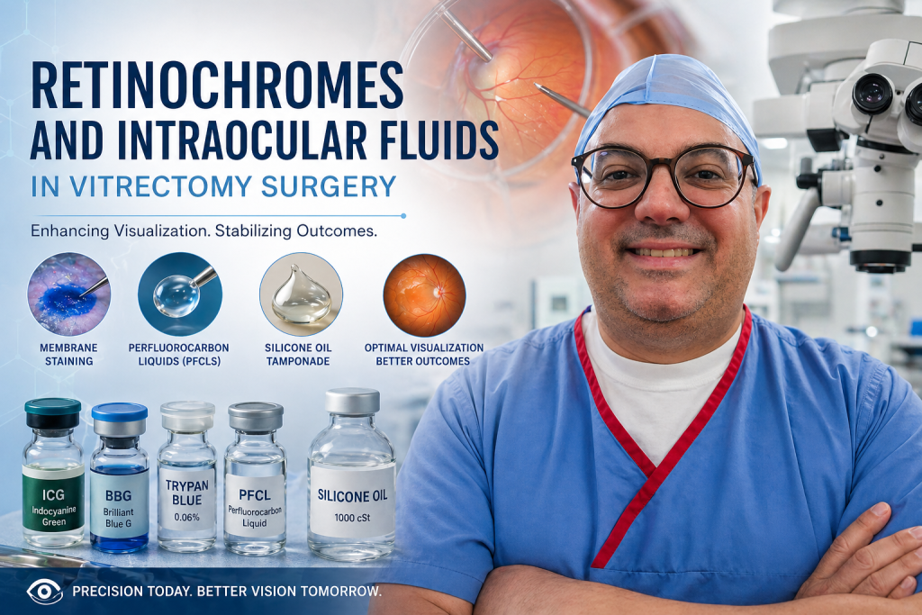
Vitrectomy surgery has undergone remarkable evolution over the past decades, becoming a cornerstone of modern vitreoretinal practice. As surgical techniques have advanced, the importance of intraoperative visualization and retinal stabilization has become increasingly evident. Two essential components that have significantly contributed to these improvements are retinochromes (vital dyes) like , Indocyanine Green, Brilliant Blue G, Trypan Blue and specialized intraocular fluids such as silicone oil and perfluorocarbon liquids (PFCLs). These agents have transformed the way ophthalmologists approach complex retinal conditions, allowing for greater precision, improved anatomical success, and more predictable visual outcomes. Understanding their mechanisms, applications, and limitations is therefore critical for optimizing surgical performance.
Retinochromes in Vitrectomy Surgery
Enhancing Visualization with Vital Dyes
Retinochromes, commonly referred to as vital dyes, play a fundamental role in enhancing intraoperative visualization. The transparent nature of intraocular structures such as the internal limiting membrane (ILM) and epiretinal membranes (ERM) presents a significant challenge during surgery. Without adequate contrast, even experienced surgeons may struggle to achieve complete and safe membrane removal. Vital dyes address this limitation by selectively staining these structures, thereby improving visibility and facilitating more controlled surgical maneuvers.
Indocyanine Green (ICG) and Its Clinical Role
Among these agents, indocyanine green (ICG) has historically been one of the most widely utilized dyes, particularly for ILM staining in macular hole surgery. Its strong affinity for the ILM allows for clear delineation of the membrane, which can significantly enhance peeling efficiency. However, concerns regarding retinal toxicity have led to a more cautious approach. Evidence suggests that high concentrations, prolonged exposure, and intense illumination may contribute to retinal pigment epithelium and ganglion cell damage. Consequently, many surgeons now employ diluted solutions and minimize exposure time to reduce these risks.
Brilliant Blue G (BBG) as a Safer Alternative
In response to the limitations of ICG, alternative dyes with improved safety profiles have been developed. Brilliant Blue G (BBG) has emerged as a preferred option for ILM staining due to its selective affinity and minimal retinal toxicity. It provides sufficient contrast without deeply penetrating retinal layers, thereby reducing the risk of structural damage. As a result, BBG is widely used in macular hole repair and epiretinal membrane surgery.
Trypan Blue and Epiretinal Membrane Staining
Trypan blue is another important dye, particularly effective in staining epiretinal membranes. It enhances the visualization of fibrocellular tissue, which is often difficult to distinguish under standard illumination. This makes it especially useful in cases involving proliferative vitreoretinopathy or complex ERM peeling, where precise identification of tissue planes is essential.
Dual and Heavy Dye Formulations
More recently, combined dye formulations have been introduced, incorporating multiple staining agents within a single solution. These dual dyes enable simultaneous visualization of different retinal layers, thereby simplifying surgical workflow and reducing operative time. In addition, heavier dye formulations have been developed to improve sedimentation over the posterior pole without requiring fluid-air exchange. These innovations contribute to more efficient and controlled surgical procedures.
Intraocular Fluids in Vitrectomy Surgery
The Importance of Fluid Dynamics in Retinal Surgery
While dyes improve visualization, intraocular fluids are essential for maintaining retinal stability and achieving anatomical success. These agents provide mechanical support, facilitate fluid displacement, and assist in retinal reattachment. Among them, perfluorocarbon liquids and silicone oil play particularly significant roles in modern vitreoretinal surgery.
Perfluorocarbon Liquids (PFCLs)
Mechanism and Physical Properties
Perfluorocarbon liquids are characterized by their high specific gravity and optical clarity. These properties allow them to exert controlled mechanical pressure on the retina, effectively flattening it against the retinal pigment epithelium. This feature is especially valuable in complex retinal detachment cases.
Clinical Applications in Complex Retinal Detachment
PFCLs are widely used in the management of giant retinal tears, advanced proliferative vitreoretinopathy, and other complex retinal detachments. By displacing subretinal fluid anteriorly and stabilizing the posterior pole, they facilitate safer membrane peeling and reduce the risk of retinal slippage during surgery.
Limitations and Surgical Considerations
Despite their advantages, PFCLs are intended for intraoperative use only and must be completely removed before concluding the procedure. Retained droplets may lead to inflammation, mechanical damage, or long-term toxicity, highlighting the importance of meticulous surgical technique.
Silicone Oil Tamponade
Properties and Types of Silicone Oil
Silicone oil serves as a long-term intraocular tamponade and is available in different viscosities, typically ranging from 1000 to 5000 centistokes. Lower viscosity oils are easier to inject and remove, whereas higher viscosity formulations are more resistant to emulsification but can be technically more challenging to handle.
Indications for Silicone Oil Use
Silicone oil is commonly indicated in complex vitreoretinal conditions, including proliferative vitreoretinopathy, diabetic tractional retinal detachment, ocular trauma, and recurrent retinal detachment. Its ability to provide prolonged support makes it particularly useful in challenging cases.
Advantages and Clinical Benefits
Unlike intraocular gases, silicone oil does not resorb spontaneously, allowing for continuous retinal support over an extended period. This is particularly beneficial for patients who are unable to maintain strict postoperative positioning or require long-term tamponade.
Complications and Management
Despite its advantages, silicone oil may lead to complications such as cataract formation, increased intraocular pressure, and emulsification. These risks necessitate careful patient selection and timely removal to optimize outcomes.
Integration of Dyes and Fluids in Surgical Practice
The combined use of retinochromes and intraocular fluids has significantly enhanced the precision and safety of vitrectomy surgery. For instance, staining the ILM with Brilliant Blue G while using PFCL to stabilize the retina allows for controlled and efficient membrane peeling, even in complex cases. Following successful retinal reattachment, silicone oil can provide sustained postoperative support. This integrated approach improves both anatomical success rates and functional visual outcomes.
Future Directions in Vitreoretinal Surgery
Ongoing research continues to refine these surgical adjuncts. New dye formulations aim to reduce toxicity while improving selectivity, and advances in tamponade agents are focused on enhancing biocompatibility and long-term stability. Emerging technologies, including heavier silicone oils and hybrid compounds, are being explored to address limitations in current treatment strategies, particularly for inferior retinal pathology.
Conclusion
The use of retinochromes and intraocular fluids in vitrectomy surgery represents a critical aspect of modern ophthalmic practice. Vital dyes such as indocyanine green, Brilliant Blue G, and trypan blue have revolutionized intraoperative visualization, while intraocular agents like perfluorocarbon liquids and silicone oil provide essential mechanical support and tamponade. A comprehensive understanding of these tools enables ophthalmologists to tailor surgical strategies to individual patient needs, ultimately improving both anatomical and functional outcomes.
علاج الحول الأنسي و الحول الوحشي و جميع أنواع الحول العامودي. نتائج رائعه ، و أثبتنا جدارتنا علي مر السنين. للحجز و الإستفسار انقر علي الرقم أو أرسل واتس أب 00962797262600
أو بإمكانك قراءه المزيد علي صفحتنا


